
Tell us what you need and our team will get back to you with pricing and product details within 2 hours.
Quickly find models, categories, and inspection systems across our product catalog.
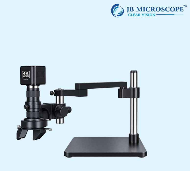
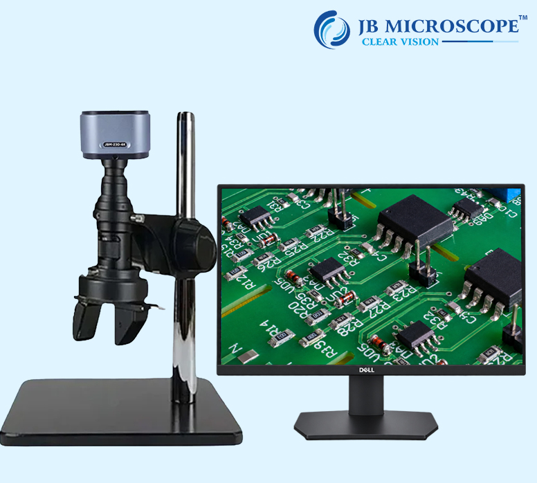
The Monozoom 3D Microscope represents a cutting-edge advancement in microscopy technology, offering a revolutionary approach to scientific exploration and observation. This sophisticated microscope is designed to provide researchers and sci...
The Monozoom 3D Microscope represents a cutting-edge advancement in microscopy technology, offering a revolutionary approach to scientific exploration and observation. This sophisticated microscope is designed to provide researchers and scientists with an immersive three-dimensional experience, allowing for unprecedented insights into the microcosms.
When observing the surface of an object with a microscope, it can be very difficult to determine the shape or topography of the surface since only a small area can be in focus at one time. This is especially true when capturing a photo with a camera. Some digital microscopes have the ability to scan an entire surface and capture each different area that comes into focus, combining them into one fully-focused image. The relative height data from each focal plane can then be used to reconstruct the surface into a 3D image that users can freely manipulate, making it easy to understand the true shape of the surface.
D.F.D. stands for Depth from Defocus. The D.F.D. method calculates three-dimensional depth information based on the amount of defocusing of two-dimensional images. When the lens is shifted vertically towards the target to capture images at different heights, the focus level of an individual point changes gradually. When a position on the target is higher, in a topographical sense, that part of the image will come into focus sooner (when moving the lens downward toward the part). When a position on the target is lower, that part of the image will come into focus as the lens is moved closer to the target. By calculating the height position of the lens when each point in the image comes into focus, a 3D image can be created. Even if perfect focus is unable to be obtained for some areas, the D.F.D. algorithm is still capable of calculating the height location of those pixels and obtaining detailed texture information.
| Parameter | Specification |
|---|---|
| Brand | JB MICROSCOPE |
| Model | JBM-HD3D |
| Zoom Ratio | 1:7 |
| Zoom Objective | 0.7X - 5X |
| Eyepiece | 0.4X |
| Working Distance | 100mm |
| Vertical Post Height | 240mm |
| Vertical Post Diameter | φ32mm |
| Microscope Body Holder | φ50mm |
| Focus Travel | 50mm |
| Base Size | 320x260x16mm (L1 Base) |
| Material | Metal |
| LED-144TP Illuminator Specification | Value |
|---|---|
| Outer Diameter | 100mm |
| Inner Diameter | 61mm |
| Wattage | 4.5W |
| Input Voltage | 90V - 264V |
| Output Voltage | DC 12V |
| Brightness Adjust | 0 - 100% |
| Working Distance | 40 - 250mm |
| Quantity of LED | 144 PCS |
| Brightness | 20000 Lux at height of 100mm |
| Color of Light | 6400K |